Magnetic Resonance is About to Get Faster, Cheaper and More Valuable

MRI Test
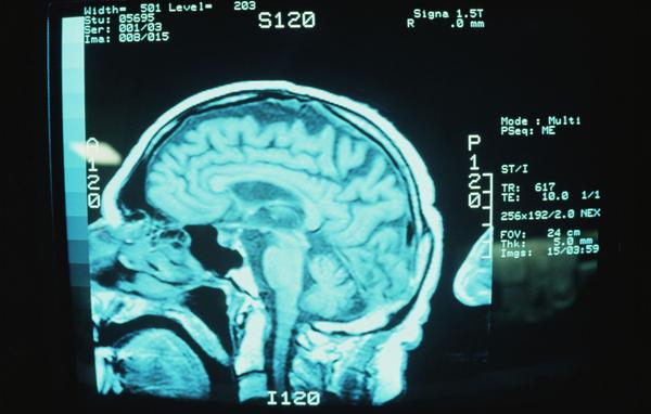
It’s been almost 40 years since the first human magnetic resonance (MR) pictures were taken. Along with decades of experience we’ve seen the development of custom sequencing and mind-boggling advances in computer processing. Yet there has been virtually no change in the length of exams.

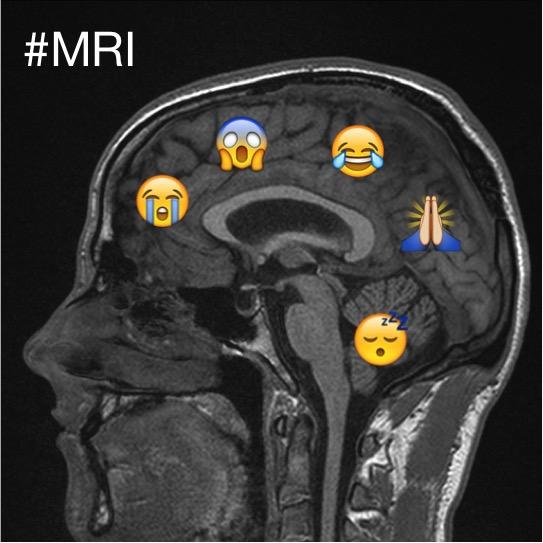
I was at a loss to explain it to my son a couple weeks ago, when he asked me why it took an hour to scan his shoulders. Before putting in the earplugs to guard against the other worldly noises of MR, he was coached by the technologist to stay as still as he could. Movement, he was told, might result in the need for a retake.

He spent an hour in that cylinder, about the same exam time for patients in the 1980s. I received a second dose of déjà vu when the summary of charges arrived. When my wife me asked me to guess, being something of a cynic, I price it at what I thought was the high-end — $2,500 maybe. I was 300% too low.

[Read more…]

‘Emotional’ brain circuit study is the first to use MRI to track depression

mri-depressionIt has been long suspected that mothers can ‘pass on’ depression to their daughters.

Now scientists have pinpointed a circuit in the brain involved in regulating emotion and mood disorders, which is handed down through the female line of families.

Researchers believe the wiring in a key brain structure, known as the corticolimbic system, may be an inherited factor contributing to depression. [Read more…]

Metamaterials boost sensitivity of MRI machines

services-mri
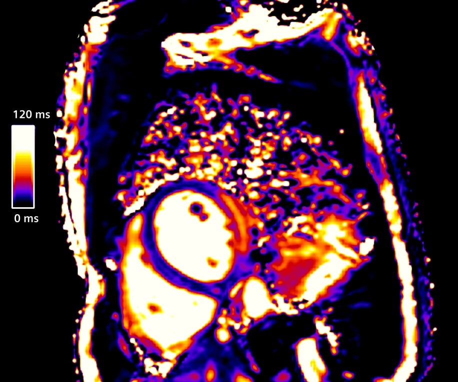
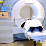
A group of researchers from Russia, Australia and the Netherlands has developed a technology that can reduce magnetic resonance imaging (MRI) scanning times by more than 50 percent, allowing hospitals to drastically increase the number of scans without changing equipment. This extraordinary leap in efficiency is achieved by placing a layer of metamaterials on the bed of the scanner, which improves the signal-to-noise ratio. The details of this experimental research are available in the current issue of Advanced Materials. This patent-pending technology is currently being co-developed by MediWise, a U.K.-based company that specializes in commercializing metamaterials for medical applications. [Read more…]

Better Brain Scans? Thank Twitter

mri-twitterGetting an MRI can be a stressful—and not just because of the physical experience of being placed into a cramped, uncomfortable scanning machine. There’s also the psychological pressure of knowing that you’re getting the procedure done in the first place because of some sort of medical necessity. To get an MRI is, in other words, entirely—understandably!—nerve-wracking. To help alleviate the stress involved in getting scanned, researchers have turned to an unlikely source of inspiration: Twitter.

In a paper to be published in the upcoming issue of the Journal of Medical Imaging and Radiation Sciences, Johnathan Hewis, who works out of Australia’s Charles Sturt University, details the process by which he and his team analyzed hundreds of MRI-related tweets over the course of a month, and used their findings to help improve the overall experience of patients slated to be scanned. A press release on the researcher’s findings explained: [Read more…]

MRI scans could predict patients at risk of major depressive disorder

mri-scanFunctional magnetic resonance imaging (fMRI) could be used to predict which patients with recovered major depressive disorder are most likely to have more depressive episodes, according to a study published today in JAMA Psychiatry.

Researchers from King’s College London and The University of Manchester, funded by the Medical Research Council, gave 64 patients who were in remission from major depressive disorder, and not on prescribed medication, fMRI scans to look for atypical connections in the brain.

During the scans the participants were asked to imagine acting badly towards their best friends and they experienced self-blaming emotions such as guilt. Over the following 14 months they were seen regularly and monitored for symptoms. At the end of the study 37 remained in remission while 27 had had a recurrence of their depression. [Read more…]

MRI scanners can steer tumor busting viruses to specific target sites within the body

mri4Scientists from the University of Sheffield have discovered MRI scanners, normally used to produce images, can steer cell-based, tumour busting therapies to specific target sites in the body.

Magnetic resonance imaging (MRI) scanners have been used since the 1980s to take detailed images inside the body – helping doctors to make a medical diagnosis and investigate the staging of a disease. [Read more…]

CT scans can damage patients DNA

CT scansCellular damage occurs when people undergo CT scans, but whether or not this causes cancer or any other health problems is unclear, a new study finds.

“The use of medical imaging for heart disease has exploded in the past decade,” study senior author Dr. Joseph Wu, director of the Stanford Cardiovascular Institute, said in a Stanford news release.

“These tests expose patients to a non-trivial amount of low-dose radiation,” Wu added. “But nobody really knows exactly what this low-dose radiation does to the patient. We now have the technology that allows us to look at very subtle, cell-level changes.”

[Read more…]

New MRI Application Provides Quantitative Information about Cardiac Tissue

myomapsMyoMaps is the world’s first MRI application able to provide quantative information regarding the composition of heart muscle tissue and represent changes in such tissues in color in the final image. This can help address so-called diffuse myocardial pathologies, in which the tissue changes are small but distributed over the whole heart. One example is the accumulation of iron deposits in the heart, which can lead to cardiac insufficiency. Siemens recently presented this solution at last year’s ESC Congress in Barcelona.
[Read more…]

White Matter Damage in Brain May Help Spot Early Alzheimer’s

brain-mri-alzheimerDamage to the brain’s white matter may be an early sign of certain types of Alzheimer’s disease, according to a new study.

Researchers used a specialized MRI technique called diffusion tensor imaging (DTI) to assess white matter in 53 people with three different types of Alzheimer’s. Some patients had atypical forms of the disorder that affect localized parts of the brain (called focal AD syndromes). These atypical forms may cause vision and language problems.

Other patients in the study had early onset Alzheimer’s, which affects several areas of the brain and interferes with thinking skills such as reasoning, planning and problem solving. This is different than late-onset Alzheimer’s, which develops after age 65 and is marked by progressive memory loss.

[Read more…]

Risks of X-rays and CT scans

mri-scanPatients are often exposed to cancer-causing radiation for little medical reason, a Consumer Reports investigation finds.

When James Duncan, M.D., a radiologist at Washington University in St. Louis, experienced intense pain in his abdomen in 2010, he rushed to a local emergency room. His doctors suspected kidney stones, but they wanted to be sure, so they ordered a CT scan. Duncan remained motionless as the machine captured a detailed, 3D image of his abdomen. He knew that the test was done when the machine stopped whirring. So he was surprised when the scanner kicked back on after a few seconds.

[Read more…]