Ted Lindsay Foundation Courage Awards

The Courage Awards annually recognize a person and their family who has been diagnosed with an Autism Spectrum Disorder and demonstrates great character and perseverance in living with the challenges of this disorder.

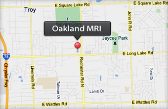
There will be two awards presented. One will recognize an individual, sponsored by Susan V. Swider and Oakland MRI. The other will acknowledge the individual and their family, sponsored by Dr. Lucia Zamorano and Michigan Brain & Spine Surgery Center. The award will be presented to the winners at the annual Ted Lindsay Foundation Celebrity Golf Outing on Monday September 12, 2022 at Detroit Golf Club, Detroit, MI.

Ted Lindsay Foundation support research and educational programs focusing on the cause and management of Autism Spectrum Disorders.

Oakland MRI Is Hiring – Clinical/Administrative Assistant

Oakland MRI is looking for Clinical/Administrative Assistant to join our team of highly dedicated professionals who are committed in providing top-quality patient care. The candidate should possess attributes that include excellent communication skills, time-management skills, self-motivation, and the ability to work independently and as a team to accomplish daily goals and objectives, as well as, treating all patients’ and co-workers’ in a professional, respectful manner.

Learn more about this opportunity

Cardiac MRI to Identify Potential Fatal Heart Disease?

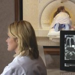
The use of MRI to determine heart function has been slow to catch on, but a study from Duke Health researchers shows that stress cardiac MRI not only diagnoses disease but can also predict which cases are potentially fatal. Results from a large, multi-center study suggest that Cardiac Magnetic Resonance, or CMR, has potential as a non-invasive, non-toxic alternative to stress echocardiograms, catheterizations and stress nuclear exams in identifying the severity of coronary artery disease. The study appears online Feb. 8 in JAMA Cardiology. “We’ve known for some time that CMR is effective at diagnosing coronary artery disease, but it’s still not commonly used and represents less than one percent of stress tests used in this country,” said senior author Robert Judd, Ph.D., co-director of the Duke Cardiovascular Magnetic Resonance Center.

“One of the impediments to broader use has been a lack of data on its predictive value — something competing technologies have,” Judd said. “Our study provides some clarity, although direct comparisons between CMR and other technologies would be definitive.” Judd and colleagues analyzed data from more than 9,000 patients who underwent CMR at seven U.S. hospitals, encompassing up to 10 years of follow-up. [Read more…]

Deadly Brain Tumours Could Be Heated Up By MRI Scanners Until They Die In Groundbreaking Treatment

Magnetic ‘metal seed’ that destroys brain tumours in ten minutes invented by British scientists

Deadly brain tumours could be removed in just ten minutes with a groundbreaking new treatment which uses MRI scanners to heat up cancer cells until they die.

The new therapy, developed by University College London, involves injecting a tiny magnetic metal ‘seed’ into the bloodstream and directing it to the site of the cancer.

The scanner is then used to heat up the metal seed which causes the cells to die in the surrounding tissue. Not only does it quickly kill cancer cells, but it saves healthy cells from the damaging effects of invasive surgery or radiotherapy.

The team at UCL has already proven it is effective in the brains of pigs and plans to move to human trials on patients with prostate cancer within the next two years with the hope it will be available for many cancers on the NHS within five years.

Launching the new technology at The Cheltenham Science Festival, Mark Lythgoe, professor of imaging at UCL, said: “The aim is to turn every MRI scanner in the world into a therapeutic device. At the moment it just take pictures.

“The simple idea is the patient goes into the MRI scanner, you locate a tumour in the brain or the prostate and then we implant a tiny magnetic particle, a little bit smaller than a grain of rice, to the site of the tumour.

“We can guide it with real precision avoiding any areas that we don’t want to go to, like the sensory motor-cortex in the brain, the area with memories.

“Once it’s in there we’re able to fire in a simple radio wave and these seeds heat up remarkably well, and kills all the cells around it. You then just guide the seed through the tumour, killing all the cells. And you can do it with real precision right up to the margins of the tumour so there is no tumour left. This is life-changing.”

Read more

A Brief History Of Medical MRI, Starting With A McDonald’s

In 1983, Robert “Bob” Kagan, a 36-year-old pathologist, set out on foot from Holy Cross Hospital in Ft. Lauderdale, Florida, looking for a place to birth an industry. He’d find it in a McDonald’s, which would become one of America’s first outpatient MRI centers.

Kagan had become convinced of the promise of magnetic resonance imaging, a now-commonplace technology that was then still experimental. The Food and Drug Administration would not approve MRI until 1984, and Medicare wouldn’t cover it until 1985. Holy Cross Hospital refused to spend the $1.2 million ($3 million in 2017 dollars) for an MRI machine. But other doctors at the hospital saw the potential, and 40 put up $25,000 each for Kagan’s new business: an MRI machine that would sit outside the hospital walls. [Read more…]

8 Unexpected Activities People Have Done in MRI Scanners for Science

In medicine, magnetic resonance imaging (MRI) uses powerful magnetic fields and radio waves to show what’s happening inside the body, producing dynamic images of our internal organs. Using similar technology that tracks blood flow, functional magnetic resonance imaging (fMRI) scans can show neuroscientists neural activity, indicating what parts of the brain light up when, for instance, a person thinks of an upsetting memory or starts craving cocaine. Both require staying within a massive MRI machine for the length of the scan.

There’s some controversy over how scientists interpret fRMI data in particular—fMRI studies are based on the idea that an increase of blood flow to a region of the brain means more cellular activity there, but that might not be a completely accurate measure, and a 2016 report found that fMRI studies may have stunning rates of false positives.

But we’re not here to talk about results. We’re here to talk about all the weird, weird things scientists have asked people to do in MRI machines so that they could look at their brains and bodies. From getting naked to going to the bathroom, people have been willing to do some unexpected activities in the name of science. Here are just a few of the oddest things that people have done in scanners at the behest of curious researchers.

1. SING OPERA

Researchers once invited world-famous opera singer Michael Volle to sing inside an MRI at the University of Freiburg in Germany. The baritone sang a piece from Richard Wagner’s opera Tannhäuser as part of a 2016 study on how the vocal tract moves during singing at different pitches and while changing volume. The study asked 11 other professional singers with different voice types to participate as well. They found that the larynx rose with a singer’s pitch, but got lower as the song got louder, and that certain factors, like how open their lips were, correlated more with how loud the singer was than how high they were singing. The scientists concluded that future research on the larynx and the physical aspects of singing should take loudness into consideration.

That study wasn’t the first to take MRI images of singers. In 2015, researchers at the University of Illinois demonstrated their technique for recording dynamic MRI imaging of speech using video of U of I speech specialist Aaron Johnson singing “If I Only Had a Brain” from The Wizard of Oz.

Read full article

Taking MRI Technology Down To Micrometer Scales

MRI Services
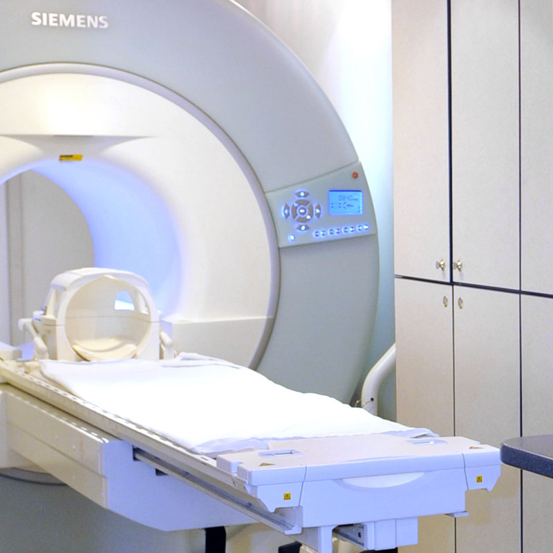
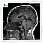
Millions of magnetic resonance imaging (MRI) scans are performed each year to diagnose health conditions and perform biomedical research. The different tissues in our bodies react to magnetic fields in varied ways, allowing images of our anatomy to be generated. But there are limits to the resolution of these images—generally, doctors can see details of organs as small as a half millimeter in size but not much smaller. Based on what the doctors see, they try to infer what is happening to cells in the tissue.

Mikhail Shapiro, assistant professor of chemical engineering, wants to make a connection between MRI images and what happens in tissues at scales as small as a single micrometer—that’s about 500 times smaller than what’s possible now.

“When you look at a splotchy MRI picture, you may want to know what’s happening in a certain dark spot,” says Shapiro, who is also a Schlinger Scholar and Heritage Medical Research Institute Investigator. “Right now, it is hard to say what’s going on at scales smaller than about half a millimeter.”

In a recent study published in the journal Nature Communications, Shapiro and his colleagues introduced a method to correlate magnetic field patterns in tissue, which occur on micrometer scales, with the larger, millimeter-scale features of MRI images. Ultimately, the method would allow doctors to interpret MRI pictures and better diagnose various conditions.

For example, medical researchers can visualize the locations of inflamed tissues in a patient’s body by using MRI to take images of immune cells called macrophages that have been labeled with magnetic iron particles. The macrophages take up iron particles injected into a patient’s bloodstream and then migrate toward sites of inflammation. Because the MRI signal is affected by the presence of these iron particles, the resulting images reveal locations of unhealthy tissue. However, the exact level of MRI contrast depends on precisely how the cells take up and store the iron particles on the micrometer scale, which cannot be seen directly in the MRI images.

The new technique could provide an understanding of how different iron distributions affect MRI contrast, and this, in turn, would provide a better idea of the scope of inflammation. The research was led by Caltech graduate students Hunter Davis and Pradeep Ramesh.

Read more at: https://phys.org/news/2018-03-mri-technology-micrometer-scales.html#jCp

Abbreviated Breast MRI May Be Additional Screening Option for Dense Breasts

Among women with dense breast tissue, for whom traditional mammograms are less effective at detecting cancer, who request additional screening after a negative mammogram, abbreviated breast MRI (AB-MR) may be a valuable cancer detection tool. In a study of 195 asymptomatic women with dense breast tissue who had a negative mammogram within the previous 11 months, AB-MR detected five additional cancers after a negative screening mammography, according to preliminary findings from a Penn Medicine team presented this week at the Radiological Society of North America meeting in Chicago.

To put this in perspective, the cancer detection rate of mammography is roughly 4 cancers in 1,000 women who have a mammogram. Digital tomosynthesis (DBT), or 3D mammography, does slightly better, detecting approximately 25 percent more cancers, or roughly 5 cancers in 1,000 women screened. Based on the preliminary results at Penn Medicine, the cancer detection rate of AB-MR screening is 25 cancers per 1,000 patients. One in eight women in the United States will develop breast cancer at some point during their life.

“Having dense breast tissue makes it more difficult to detect a cancer on a mammogram,” said the study’s lead author, Susan Weinstein, MD, an associate professor of Radiology and the director of breast MRI at Penn Medicine. “Based on the literature and our results, women with dense breast tissue who desire supplemental screening, these results suggest that AB-MR may be a better option than other supplemental screening tests such as whole breast ultrasound.

The most common exam offered for asymptomatic patients seeking supplemental screening is a whole breast screening ultrasound examination. However, screening ultrasound examinations have higher rates of false positives, meaning more cases of positive screenings where no cancer is present.

Based on the results from Penn’s study, the AB-MR may be a better option. American Cancer Society guidelines currently recommend a full breast MRI, not an AB-MR, in women who, based on family history of breast or ovarian cancer and/or previous treatment for Hodgkin disease, have a 20 to 25 percent or greater lifetime risk of breast cancer.

In addition to Weinstein, additional authors include Mitchell D. Schnall, Elizabeth S. McDonald, Alice Chong, and Emily F. Conant.

Oakland MRI Is Hiring!

Oakland MRI is looking for Clinical/Administrative Assistant to join our team of highly dedicated professionals who are committed in providing top-quality patient care. The candidate should possess attributes that include excellent communication skills, time-management skills, self-motivation, and the ability to work independently and as a team to accomplish daily goals and objectives, as well as, treating all patients’ and co-workers’ in a professional, respectful manner.

Learn more about this opportunity

Routine Mammography Not Necessary If Used Breast MRI

3d-breast-mri
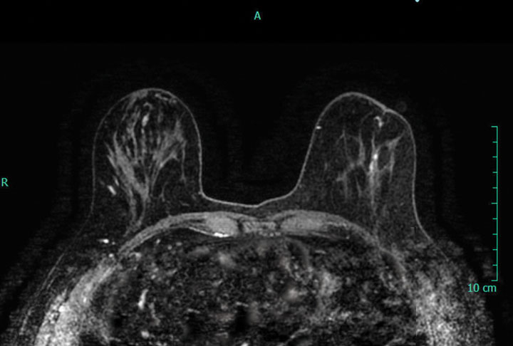
Adding screening mammography to annual screening MR imaging for women at high risk for breast cancer does not increase cancer detection rates, according to a study published in Radiology.

Researchers from Canada performed a retrospective review to evaluate the value of mammography in detecting breast cancer in high-risk women undergoing screening breast MR imaging.

The researchers evaluated 3,934 screening studies (1,977 screening MR imaging examinations and 1,957 screening mammograms) performed in 1,249 high-risk women. The performance measures included recall and cancer detection rates, sensitivity, specificity, and positive predictive values were calculated for both mammography and MR imaging. [Read more…]