 Suspected transient ischemic attacks (TIAs) – Noninvasive cervical vessel imaging
- Transient ischemic symptoms – Neuroimaging within 24 hours
- Steno-occlusive disease – CT angiography or MR angiography of intracranial vasculature
The Bottom Line: MRI remains preferred over CT for imaging patients with suspected TIAs because it can provide insight into whether a stroke has occurred.
Unchanged: In patients with suspected TIAs, noninvasive imaging of cervical vessels is indicated. In those with transient ischemic neurologic symptoms, neuroimaging is recommended within 24 hours of symptom onset or as soon as possible in cases of delayed presentation. MRI is preferred over CT.
Revised: In cases of known steno-occlusive disease, CT angiography or MR angiography of intracranial vasculature is recommended to assess for proximal intracranial stenosis and/or occlusion. Catheter angiography is necessary to confirm diagnosis and assess stenosis severity.
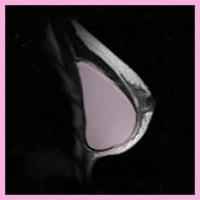 NBC’s Nightly News With Brian Williams, NBC’s Chief Medical Expert Dr. Nancy Snyderman reviewed a new US Food and Drug Administration (FDA) report in regard to silicone gel-filled breast implants. The report indicated that women should undergo surveillance with a breast MRI 3 years after implantation, and subsequently every 2 years. The FDA has recommended surveillance MRI for silent rupture since 2006, when silicone implants came back on the market for cosmetic augmentation.
 Three-fourths of patients with Alzheimer’s disease or frontal-lobe degeneration had MRI-detected biomarker levels that correlated with the diagnoses, suggesting MRI has potential as a screening tool for the conditions, investigators reported.