Study shows MRIs are safe for patients with wide variety of pacemakers, defibrillators

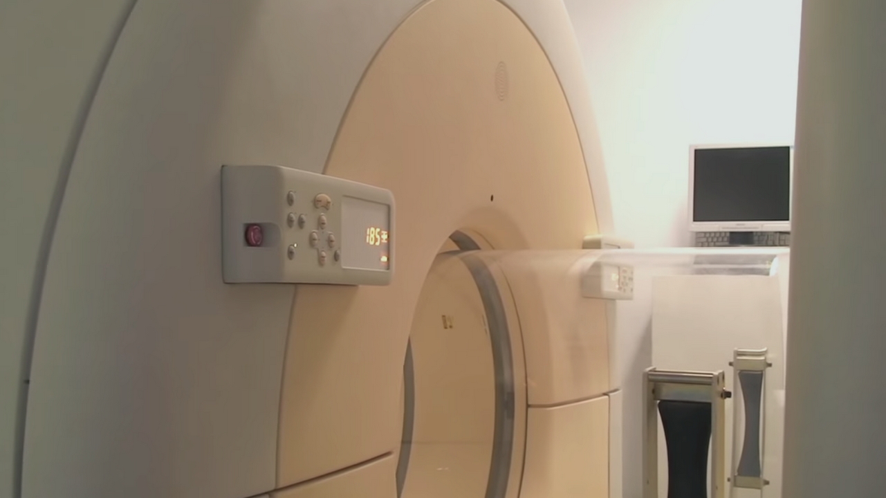
Magnetic resonance imaging appears to be safe for patients with cardiac implantable electronic devices, even for chest imaging, according to a new study by researchers from the Intermountain Medical Center Heart Institute in Salt Lake City.

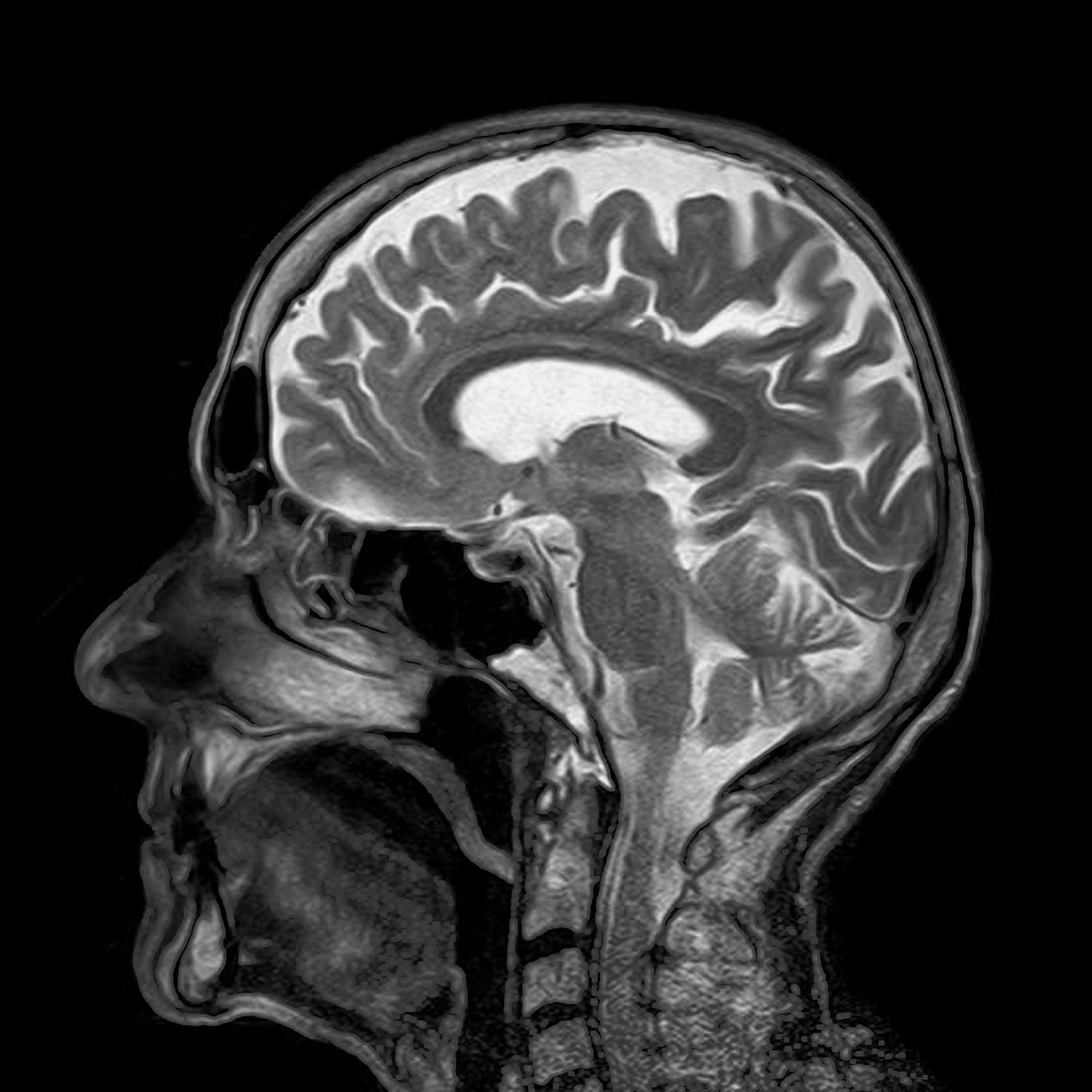
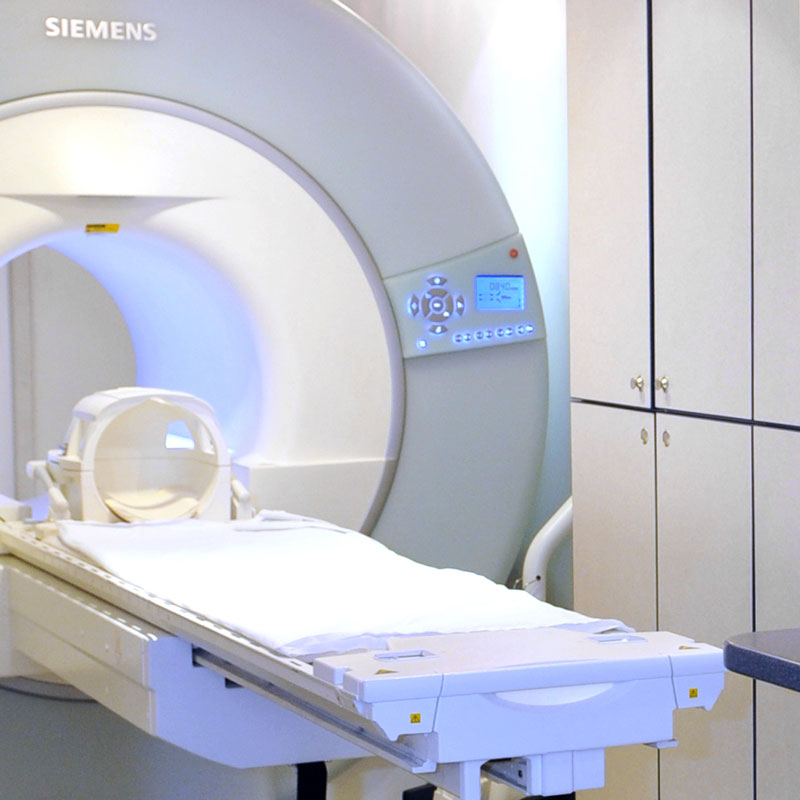
In the past, MRIs have been considered dangerous for people who have the devices. However, the new study, “Real World MRI Experience with Non-Conditional and Conditional Cardiac Rhythm Devices After MagnaSafe,” published in the Journal of Clinical Electrophysiology, found that MRI imaging can be safely performed on patients with devices.

“Magnetic resonance imaging has become very popular,” said Jeffrey L. Anderson, MD, senior study author and cardiologist at the Intermountain Medical Center Heart Institute. “It’s excellent for looking at soft tissue changes. But it involves very high-strength magnetic fields, which means if a patient has any implanted metal devices containing iron, it could potentially cause harm.” [Read more…]

Using MRI Research to Help Understand The Brain

Researchers and clinicians now have a new tool to help them further their understanding of disease and the complexities of the human body.

Scientists have made incredible strides in understanding the world around us. They’ve shot astronauts into space, explored deep beneath the ocean and created powerful computers that fit in our pockets. The cosmos might be well-mapped, but they’re still searching for a cure for cancer and just beginning to understand the complex connections in the brain.

Research is critical to breaking through these medical frontiers and bringing life-changing answers to millions of people around the world. MRI is a critical tool that is helping researchers better understand the human body non-invasively but it’s still a fairly young technology. Now researchers and clinicians have a new tool to help them further their understanding of disease and the complexities of the human body. [Read more…]

Researchers discover MRI can measure kidney scarring and predict future kidney function

Researchers from St. Michael’s Hospital have made what are believed to be two world first discoveries: an MRI can measure kidney damage and can predict future kidney function within one year while avoiding needle biopsies.

The researchers used a specific magnetic resonance imaging test called an elastogram to measure kidney scarring in 17 people who had kidney transplants, according to the study published online today in the Clinical Journal of the American Society of Nephrology.

An elastogram maps the stiffness of tissue using MRI to determine the presence of scarring, according to Dr. Anish Kirpalani, the study’s lead author, a radiologist and a scientist in the Li
Ka Shing Knowledge Institute of St. Michael’s.
[Read more…]

MRI scan that can predict stroke risk has ‘promise to save lives’

A new type of MRI scan can predict the risk of having a stroke, researchers have said in a study.

The non-invasive technique, developed by scientists at the University of Oxford, predicts whether plaques in the carotid arteries are rich in cholesterol and therefore more likely to cause a stroke.

Carotid arteries supply the brain with blood. The rupture of fatty plaques can block them and possibly starve the brain of oxygen, causing potentially debilitating and life-threatening strokes.

[Read more…]

Multicolor MRIs could aid disease detection

Researchers at Case Western Reserve University School of Medicine have developed a method that could make magnetic resonance imaging—MRI—multicolor. Current MRI techniques rely on a single contrast agent injected into a patient’s veins to vivify images. The new method uses two at once, which could allow doctors to map multiple characteristics of a patient’s internal organs in a single MRI. The strategy could serve as a research tool and even aid disease diagnosis.

“The method we developed enables, for the first time, the simultaneous detection of two different MRI contrast agents,” said Chris Flask, PhD, Associate Professor of Radiology, Biomedical Engineering, and Pediatrics, and Director of the Imaging Resource Core at Case Western Reserve University School of Medicine. Two contrast agents could include one specifically targeting diseased tissue, and one designed to show healthy tissue, for example. The new method would enable immediate comparisons of how each agent distributes in the same patient. [Read more…]

Does widespread pain stem from the brain?

CT scansPain is the most common reason people seek medical care, according to the National Institutes of Health.

“Sometimes we can easily pinpoint what is causing a person pain,” says Richard Harris, Ph.D., associate professor of anesthesiology and rheumatology at Michigan Medicine. “But, there are still 1 in 5 Americans who suffer from persistent pain that is not easily identifiable.”

Whenever someone experiences pain, they often think about how intense the pain is—but rarely do they also consider how widespread the pain is.

Harris is the senior author on a new study, published in Pain, that sought to find what underlies widespread pain. [Read more…]

MRI device could bridge neuro-technologies for medical diagnostics, increase safety

A technology being developed at Purdue University could provide an affordable, smart, self-learning device that, when placed into existing MRI machines could allow medical professionals to monitor patients more effectively and safely, by performing concurrent medical imaging and recording for diagnostic purposes.

Purdue researchers recently presented their findings, “Multimodal Imaging: MR-Compatible, Gradient Artifact free, Wireless recording system integrated with MR-scanner for Simultaneous EEG and fMRI acquisition,” at the International Society for Magnetic Resonance in Medicine conference in Honolulu, Hawaii. The article also received an ISMRM magna cum laude merit award, and power-pitch highlight, for highly-rated scientific merits. [Read more…]

Who Needs a 3D Mammogram and Benefits of 3D Breast MRI’s

Women in general can be considered for a 3D mammogram if they are being screened for breast cancer. However, women with dense tissue in their breast should not consider anything else, but 3D imaging. The ability to view the entire breast is far more difficult with dense tissue. Therefore, suspicious tissue or masses could easily be missed, reducing the opportunity of early detection.

The recommended age for women to start getting annual mammograms has changed over the years. But, many medical professionals believe that it should start at 40 to get a good baseline, making it easier to detect abnormalities and breast cancer. [Read more…]

The Benefits of a Breast MRI – Are they Safe?

When it comes to diagnosing breast cancer, a breast MRI is relatively new, but is becoming one of the most dependable tests for detecting abnormalities in the breast. It uses a powerful magnet to produce hundreds of images, using gadolinium, a non-radioactive contrast. Although, the contrast isn’t always used, especially for those who have a sensitivity to iodine.

Your doctor might ask for a breast MRI in addition to other tests, such as an ultrasound. But, the MRI has some advantages over other tests. [Read more…]

How Does 3D Mammography Differ from 2D?

Technology progression is constant, and the tools for early breast cancer detection are always improving. Women going in for breast cancer screening today could experience 2D or 3D imaging. Both are good. But, there are some differences…

  • Technology – X-ray technology is used with mammograms to generate an image of the breast. In 2D mammography, an x-ray arm shoots images of the breast, but can only generate a flat image. With 3D mammography, it can provide “slices” of images, so there are more images available to evaluate.
  • Accuracy – Because of the technology of 3D is creating far more images than the 2D technology cannot create, accuracy is greatly improved with 3D imaging. There are fewer false-positive results with 3D as well, because of the ability to view more of the tissue more clearly. This helps eliminate unnecessary repeat tests.
  • Clarity – Women with dense tissue in their breasts have been at a disadvantage with 2D imaging, because the density makes it more difficult to get clear pictures when it’s a flat image. With 3D, the clarity significantly improves, due to the ability to take images in “slices” and reconstruct the breast for imaging.
  • Safety – Exposure to radiation should be kept to a minimum, of course. But, the difference in the amount of radiation between 2D and 3D is the same. The difference is that with 3D mammography, the need for repeat testing is far less, because the clarity and accuracy is better than it is with 2D imaging.

So, there are differences between 2D and 3D mammography. However, both are still a good option for viewing the breast tissue. Your doctor will be able to help you decide which one would be best for you. But, there are reasons why 3D might be better for some women.

To schedule Breast MRI exam please call us at (248) 740-0777.