Magnetic resonance imaging (MRI) is a non-invasive test that uses a magnetic field and pulses of radio wave energy to make pictures of organs and structures inside the body. In many cases MRI gives different information about structures in the body than can be seen with an X-ray, ultrasound, or computed tomography (CT) scan. MRI also may show problems that cannot be seen with other imaging methods.
Common uses of MRI
MRI is often used to study the knee, ankle, foot, shoulder, elbow, wrist and hand. MRI is frequently used for imaging of the musculoskeletal system. It is also a highly accurate method for evaluation of soft tissue structures such as tendons and ligaments, which are seen in great detail. Even subtle injuries are easily detected. MRI is used for the diagnosis of spinal problems including spinal stenosis, spinal tumors and disc herniation. MRI is also used for imaging of cancer, functional disorders and can be used for examination of the male and female reproductive systems.
Doctors use MRI for diagnosing coronary artery disease and other heart problems. MRI imaging of the heart can help to examine the size and thickness of the chambers of the heart and determine the extent of damage caused by a heart attack or heart disease.
Now available at Oakland MRI – A completely new MRI experience

One size fits all, a completely new MRI experience. Open MRI technology offers more headroom, more legroom and more elbowroom.
How MRI Works?
MRI is short for Magnetic Resonance Imaging. It is a procedure used in hospitals to scan patients and determine the severity of certain injuries. An MRI machine uses a magnetic field and radio waves to create detailed images of the body. Common reasons people go in to get an M.R.I. are for a sprained ankle or back pain. A strong magnetic field is created by passing an electric current through the wire loops. While this is happening, other coils in the magnet send and receive radio waves. This triggers protons in the body to align themselves. Once aligned, radio waves are absorbed by the protons, which stimulate spinning. Energy is released after “exciting” the molecules, which in turn emits energy signals that are picked up by the coil. This information is then sent to a computer which processes all the signals and generates it into an image. The final product is a 3-D image representation of the area being examined.
We will be happy to answer any questions or discuss any concerns you may have.
MRI Scan
If you have ever seen an MRI machine, you know that the basic design used in most is a giant cube. The cube in a typical system might be 7 feet tall by 7 feet wide by 10 feet long (2 m by 2 m by 3 m), although new models are rapidly shrinking. There is a horizontal tube running through the magnet from front to back. This tube is known as the bore of the magnet.
The patient, lying on his or her back, slides into the bore on a special table. Whether or not the patient goes in head first or feet first, as well as how far in the magnet they will go, is determined by the type of exam to be performed. MRI scanners vary in size and shape, and newer models have some degree of openness around the sides, but the basic design is the same. Once the body part to be scanned is in the exact center or isocenter of the magnetic field, the scan can begin.
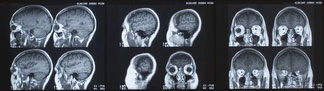
In conjunction with radio wave pulses of energy, the MRI scanner can pick out a very small point inside the patient’s body and ask it, essentially, “What type of tissue are you?” The point might be a cube that is half a millimeter on each side. The MRI system goes through the patient’s body point by point, building up a 2-D or 3-D map of tissue types. It then integrates all of this information together to create 2-D images or 3-D models.
MRI provides an unparalleled view inside the human body. The level of detail we can see is extraordinary compared with any other imaging modality. MRI is the method of choice for the diagnosis of many types of injuries and conditions because of the incredible ability to tailor the exam to the particular medical question being asked. By changing exam parameters, the MRI system can cause tissues in the body to take on different appearances. This is very helpful to the radiologist (who reads the MRI) in determining if something seen is normal or not. We know that when we do “A,” normal tissue will look like “B” — if it doesn’t, there might be an abnormality. MRI systems can also image flowing blood in virtually any part of the body. This allows us to perform studies that show the arterial system in the body, but not the tissue around it. In many cases, the MRI system can do this without a contrast injection, which is required in vascular radiology.