 Researchers and clinicians now have a new tool to help them further their understanding of disease and the complexities of the human body.
Scientists have made incredible strides in understanding the world around us. They’ve shot astronauts into space, explored deep beneath the ocean and created powerful computers that fit in our pockets. The cosmos might be well-mapped, but they’re still searching for a cure for cancer and just beginning to understand the complex connections in the brain.
Research is critical to breaking through these medical frontiers and bringing life-changing answers to millions of people around the world. MRI is a critical tool that is helping researchers better understand the human body non-invasively but it’s still a fairly young technology. Now researchers and clinicians have a new tool to help them further their understanding of disease and the complexities of the human body.
Scientists at the University of Wisconsin – Madison are using new MRI technology that can enable them to see the brain better than ever before to conduct research that will help them understand disease and how to treat it. The new SIGNA Premier, GE Healthcare’s ultra-premium 3.0T MR system, is the result of a collaboration between research institutions around the world working to design new imaging tools, particularly to aid researchers in the detection of biomarkers for the potential diagnosis of mild Traumatic Brain Injury.
Hover over the images to learn more about the technology behind using MRI to understand the brain.
Getting to the white matter
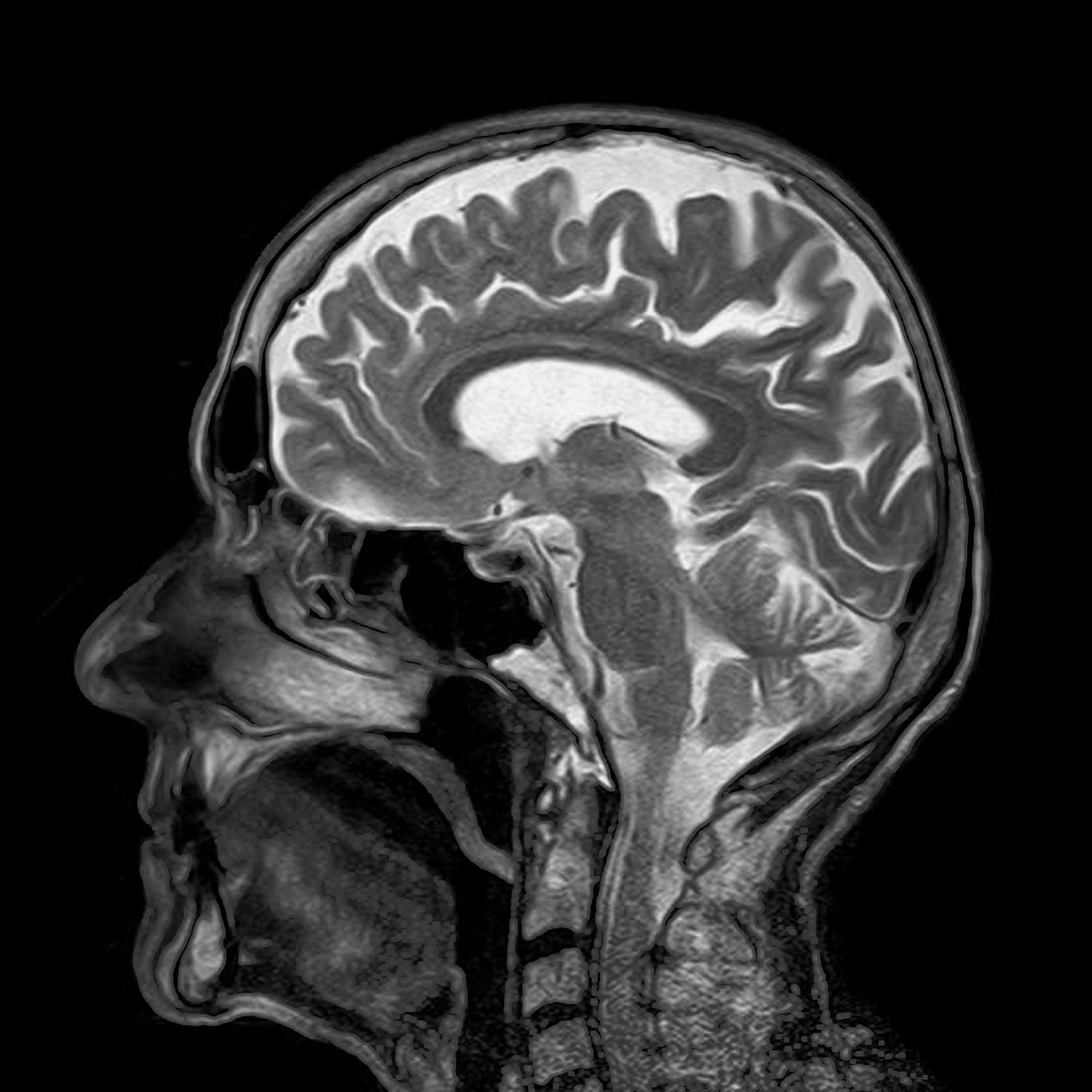
Dr. Alan McMillan and Dr. Samuel Hurley, researchers in the Department of Radiology at the University of Wisconsin, have been using SIGNA Premier in neurological research similar to the Human Connectome Project, a project to construct a map of the complete structural and functional neural connections in the brain.
Dr. McMillan and Dr. Hurley are using the SIGNA Premier in a research setting to help develop quantitative MRI methods that measure white matter in the brain throughout disease progression. They are pushing the system to its limits to get more signal and better spatial resolution, which helps to identify smaller neural tracks.
“So far we’ve been really pleased,” Dr. Hurley says. “We can set up the MR protocols we need and run them through existing Human Connectome Project image analysis pipelines. And we’re getting results that look as good or better than images we’ve seen previously.”
With visibility of the smallest details, researchers can see more than ever before, including white matter and “crossing fibers” in the brain. Dr. Hurley’s research measures the physical connections between brain regions, where white matter acts like wiring that connects the different regions of the brain together, then connects the brain to the body through the spinal cord.
“That wiring is not neat and organized. It is very complex and has lots of crossing pathways,” he says. “So to be able to get an accurate picture of how brain regions talk to each other and how they’re wired together, we need to be able to see those crossing fibers and see where these connections are going.”
SIGNA Premier allows their research team to see the fine structures of the brain and compare that data to existing databases of healthy subjects, which will likely add valuable information to further neurological research. For Dr. Hurley, this type of imaging is critical in assisting with their understanding how the body – and especially the brain – work.
“We’re getting a much more realistic view of how the brain connections are wired together and it’s enabling us to noninvasively measure the brain,” he says. “We’re pushing the imaging technology where we can get a map of the brain connections and a map of the brain wiring that appears to be the most accurate we’ve seen to date.”
Dr. Scott Reeder, professor, Vice Chair of Research, and Chief of MRI at the University of Wisconsin-Madison, has been using SIGNA Premier in his research on oncology and liver disease.
“The high performance of the system is going to allow us to not only push our current research applications to new levels in terms of spatial resolution, scan time, and also signal to noise performance, but it may offer new applications that couldn’t be done before,” he says.
The best way to understand improving signal to noise ratio is to compare it to taking a photograph of a lit candle in a dark room. Typically, with an ordinary camera, the result would be a poor image due to limited light coming from the candle flame (or in the case of an MRI, the signal) as pictured on the left in the image above. However, with a very good camera, the image that can be captured makes shapes and boundaries clearer, as seen in the image on the right. This can help push the boundaries of MRI research.
The go-to system for neurological research
SIGNA Premier is the result of a four-year collaboration with the National Football League (NFL) and research institutions around the world working to design new imaging tools, particularly to aid researchers in the detection of biomarkers for the potential diagnosis of mild Traumatic Brain Injury. SIGNA Premier delivers a new level of clinical performance with additional research-focused capabilities, especially for neurology research.
With growing reports of mild traumatic brain injury in athletes, there’s continued pressure to understand the brain so we can treat and prevent these kinds of injuries.
“SIGNA Premier is going to be our go-to neuroimaging system for many of our clinical and research applications,” says Dr. Reeder. “It is hoped that it will improve our understanding of diseases like chronic traumatic encephalopathy and mild traumatic brain injury. This is the best tool we have for performing neuroimaging exams on these patients.”
Wide bore. No limits.
SIGNA Premier features a wide bore that accommodates larger patients and those who suffer from claustrophobia. It includes digital radiofrequency transmit-and-receive architecture, enabling researchers to acquire patient data from multiple high channel-density surface coils all at once. SIGNA Premier is powered by a SuperG gradient coil, which provides outstanding performance and superb stability, and features GE Healthcare’s newest short-bore, high-homogeneity 3.0T superconductive magnet.
Historically, wide bore systems compromised image quality and research trials discouraged their use because they couldn’t meet their needs. But SIGNA Premier’s 70-centimeter wide bore doesn’t hamper researchers’ high-performance capabilities.
“It’s really cool that there’s a new wide bore system that doesn’t have any performance limitations in comparison to other systems,” says Dr. McMillan. “This system is by far and away, the best performing system that we have here at our institution, in terms of the latest development in the RF coils and the overall gradient system.”